The eyes are wonderful sensory organs. They help people learn about the world in which they live. Eyes see all sorts of things – big or small, near or far, smooth or textured, colors and dimensions. The eyes have many parts – all of which must function in order to see properly.
Inside the Eye
In addition to the many sections of the eyeball itself, muscles are attached to the outer walls of the eyeball. The eye muscles are attached to the eyes in order that we can move our eyes. The interactive diagram shows these main parts. If anything goes wrong, such as from diabetic eye disease, an individual might not be able to see as well.
A Complete Picture
Visual information from the retina travels from the eye to the brain via the optic nerve. Because eyes see from slightly different positions, the brain must mix the two images it receives to get a complete picture.
What we think of as seeing is the result of a series of events that occur between the eye, the brain, and the outside world. Light reflected from an object passes through the cornea of the eye, moves through the lens which focuses it, and then reaches the retina at the very back where it meets with a thin layer of color-sensitive cells called the rods and cones. Because the light criss-crosses while going through the cornea, the retina “sees” the image upside down. The brain then “reads” the image right-side up.
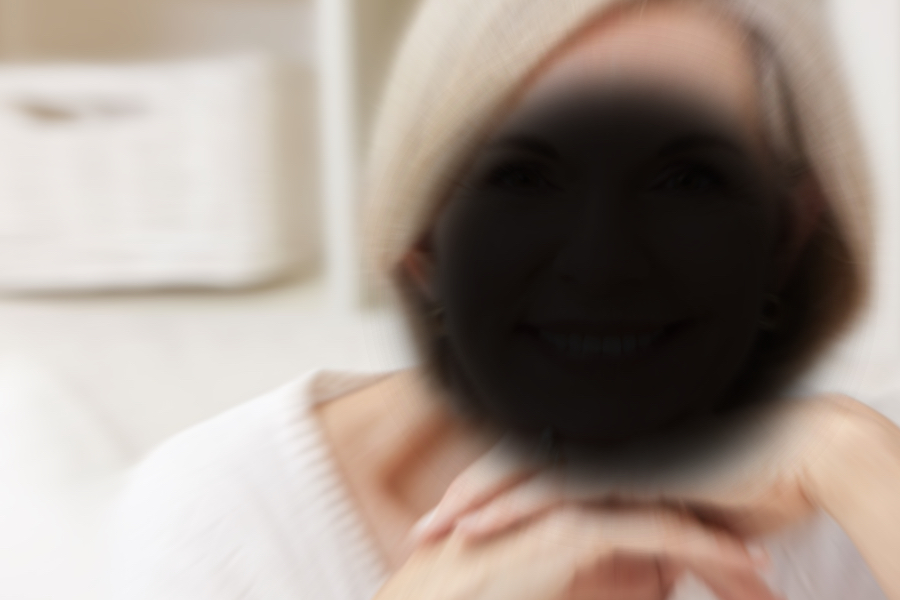
Eye diseases and conditions such as age-related macular degeneration, diabetes-related eye disease, glaucomam can cause permanent vision loss or blindness if not diagnosed and treated early. That’s why Prevent Blindness recommends that everyone receive a comprehensive eye exam through dilated pupils regularly as recommended by your eye doctor.
Simulated vision loss from diabetes-related retinopathy


Simulated vision loss from age-related macular degeneration


Simulated vision loss from cataract


Simulated vision loss from glaucoma


